
Reviews

Fractional picosecond laser-induced optical breakdown in dermatology: from controlled microinjury and molecular repair pathways to clinical endpoints and post-procedural care
Publisher's note
All claims expressed in this article are solely those of the authors and do not necessarily represent those of their affiliated organizations, or those of the publisher, the editors and the reviewers. Any product that may be evaluated in this article or claim that may be made by its manufacturer is not guaranteed or endorsed by the publisher.
Published: 31 August 2026
0
Views
0
Downloads

Authors

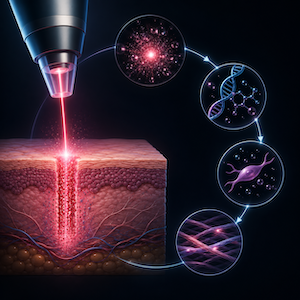
Fractional picosecond lasers have expanded the clinical use of ultrashort-pulse devices from tattoo and pigment removal to non-ablative rejuvenation, scar remodeling, enlarged pores, dyschromia and selected photoaging indications. Their distinctive tissue effect is not conventional fractional coagulation or ablation, but focal photomechanical disruption mediated by laser-induced optical breakdown (LIOB) and associated laser-induced cavitation (LIC), with vacuolization in epidermal and dermal tissue planes. This review synthesizes the translational evidence linking fractional optical delivery systems, histologic microinjury patterns, non-invasive imaging correlates, molecular repair pathways, clinical endpoints, safety across skin types and post-procedural care. PubMed-indexed literature was reviewed using search blocks for fractional picosecond lasers, LIOB/LIC, microlens and diffractive optical systems, optical coherence tomography (OCT), reflectance confocal microscopy (RCM), multiphoton and harmonic generation microscopy (HGM), wound healing, barrier repair, photoprotection and post-inflammatory hyperpigmentation (PIH). Current evidence shows that fractional picosecond systems can generate epidermal and dermal vacuoles, shockwave-associated optical signatures, melanin-dependent thresholds and remodeling responses involving matrix metalloproteinases (MMP), collagens, heat shock proteins (HSPs), cytokines, chemokines and barrier-related proteins. Clinical evidence is strongest for atrophic acne scars, photoaging, pores and selected non-acne scars, with increasing data in Asian and darker skin phototypes. However, the extent to which clinical outcomes are directly attributable to LIOB rather than combined photomechanical, photoacoustic and limited photothermal effects remains incompletely defined. Evidence-based aftercare remains underdeveloped. Conservative barrier support, strict photoprotection and avoidance of irritants are currently the most defensible post-procedural principles.

Downloads

Download data is not yet available.

Citations

1. Wu DC, Goldman MP, Wat H, Chan HHL. A systematic review of picosecond laser in dermatology: evidence and recommendations. Lasers Surg Med 2021;53:9-49.
2. Zhou Y, Hamblin MR, Wen X. An update on fractional picosecond laser treatment: histology and clinical applications. Lasers Med Sci 2023;38:45.
3. Freedman J, Kaufman J, Metelitsa A, Green J. Picosecond lasers: the next generation of short-pulsed lasers. Semin Cutan Med Surg 2014;33:164-8.
4. Saluja R, Gentile RD. Picosecond laser: tattoos and skin rejuvenation. Facial Plast Surg Clin North Am 2020;28:87-100.
5. Brauer JA, Kazlouskaya V, Alabdulrazzaq H, et al. Use of a picosecond pulse duration laser with specialized optic for treatment of facial acne scarring. JAMA Dermatol 2015;151:278-84.
6. Weiss RA, McDaniel DH, Weiss MA, et al. Safety and efficacy of a novel diffractive lens array using a picosecond 755 nm alexandrite laser for treatment of wrinkles. Lasers Surg Med 2017;49:40-4.
7. Wat H, Shek SYN, Yeung CK, Chan HHL. Efficacy and safety of picosecond 755-nm alexandrite laser with diffractive lens array for non-ablative rejuvenation in Chinese skin. Lasers Surg Med 2019;51:562-8.
8. Yu W, Zhu J, Yu W, et al. Three-year results of facial photoaging in Asian patients after alexandrite 755 nm picosecond laser with diffractive lens array: a split-face, single-blinded, randomized controlled comparison. Lasers Surg Med 2021;53:1208-17.
9. Lee CH, Jin EM, Seo HS, et al. Efficacy and safety of treatment with fractional 1,064-nm picosecond laser with diffractive optic element for wrinkles and acne scars: a clinical study. Ann Dermatol 2021;33:254-62.
10. Li J, Duan F, Kuang J. Fractional picosecond laser for atrophic acne scars: a meta-analysis. J Cosmet Dermatol 2023;22:2205-17.
11. Dai R, Cao Y, Su Y, Cai S. Comparison of 1064-nm Nd:YAG picosecond laser using fractional micro-lens array vs. ablative fractional 2940-nm Er:YAG laser for atrophic acne scar in Asians: a 20-week prospective, randomized, split-face, controlled pilot study. Front Med (Lausanne) 2023;10:1248831.
12. Yuan Y, He Y, Fang J, et al. Comparison of the fractionated Nd:YAG 1064-nm picosecond laser with holographic optics and the fractional CO2 laser in atrophic acne scar treatment: a prospective, randomized, split-face study. Int J Dermatol 2025;64:131-8.
13. Zhao S, Wang M, Shi C, et al. Clinical and histologic changes after 755-nm picosecond laser with a novel platinum focus lens array in the treatment of photo-aged skin. Lasers Med Sci 2025;40:320.
14. Habbema L, Verhagen R, Van Hal R, et al. Minimally invasive non-thermal laser technology using laser-induced optical breakdown for skin rejuvenation. J Biophotonics 2012;5:194-9.
15. Kim H, Hwang JK, Choi J, Kang HW. Dependence of laser-induced optical breakdown on skin type during 1064 nm picosecond laser treatment. J Biophotonics 2021;14:e202100129.
16. Lee HC, Childs J, Chung HJ, et al. Pattern analysis of 532- and 1,064-nm picosecond-domain laser-induced immediate tissue reactions in ex vivo pigmented micropig skin. Sci Rep 2019;9:4326.
17. Agrez V, Mur J, Petelin J, Petkovsek R. Near threshold nucleation and growth of cavitation bubbles generated with a picosecond laser. Ultrason Sonochem 2023;92:106243.
18. Linz N, Freidank S, Liang XX, Vogel A. Laser-induced plasma formation and cavitation in water: from nanoeffects to extreme states of matter. Rep Prog Phys 2025;88:086601.
19. Kim H, Hwang JK, Jung M, et al. Laser-induced optical breakdown effects of micro-lens arrays and diffractive optical elements on ex vivo porcine skin after 1064 nm picosecond laser irradiation. Biomed Opt Express 2020;11:7028-40.
20. Choi J, Duc TM, Kim H, et al. Diffractive micro-lens array for uniform and selective picosecond laser treatment. Biomed Opt Express 2023;14:2347-60.
21. Tanghetti E, Jennings J. A comparative study with a 755 nm picosecond alexandrite laser with a diffractive lens array and a 532 nm/1064 nm Nd:YAG with a holographic optic. Lasers Surg Med 2018;50:37-44.
22. Bernstein EF, Schomacker KT, Basilavecchio LD, et al. Treatment of acne scarring with a novel fractionated, dual-wavelength, picosecond-domain laser incorporating a holographic beam-splitter. Lasers Surg Med 2017;49:796-802.
23. Rhee YH, Park BC, Jung JY, et al. Effect of fractional picosecond laser therapy using a diffractive optical lens on histological tissue reaction. J Cosmet Laser Ther 2024;26:54-60.
24. Han HS, Hong JK, Park SJ, et al. A randomized, prospective, split-face pilot study to evaluate the safety and efficacy of 532-nm and 1,064-nm picosecond-domain Nd:YAG lasers using a diffractive optical element for non-ablative skin rejuvenation: clinical and histological evaluation. Ann Dermatol 2023;35:71-8.
25. Balu M, Lentsch G, Korta DZ, et al. In vivo multiphoton-microscopy of picosecond-laser-induced optical breakdown in human skin. Lasers Surg Med 2017;49:555-62.
26. Jacobsen K, Ortner VK, Fredman GL, et al. Melanin-dependent tissue interactions induced by a 755-nm picosecond-domain laser: complementary visualization by optical imaging and histology. Lasers Med Sci 2023;38:160.
27. Chang YS, Yang TH, Li CN. Histology changes of in vivo human skin after treatment with fractional 1064 nm Nd:YAG picosecond laser in different energy settings. Lasers Med Sci 2022;37:2271-8.
28. Yeh YT, Peng JH, Peng P. Histology of ex vivo skin after treatment with fractionated picosecond Nd:YAG laser in high and low-energy settings. J Cosmet Laser Ther 2020;22:43-8.
29. Wang CC. In vivo characterization of the threshold of laser-induced optical breakdown of a fractional 1064 nm Nd:YAG picosecond laser by optical coherence tomography: a step forward to precision laser therapy. J Cosmet Dermatol 2022;21:3817-20.
30. Rojas-Orrego M, Carreno N, Feuerhake T, Navarrete-Dechent C. In vivo evaluation of laser-induced optical breakdown by 1064-nm Nd:YAG fractional picosecond laser with reflectance confocal microscopy and precise histopathologic correlation. Lasers Surg Med 2024;56:650-6.
31. Liu C, Wu PJ, Chia SH, et al. Characterization of picosecond laser-induced optical breakdown using harmonic generation microscopy. Lasers Surg Med 2023;55:561-7.
32. Guida S, Fusano M, Pellacani G, Bencini PL. Fractional 1064 nm picosecond laser and skin photoaging: in vivo evaluation of treatment effects with reflectance confocal microscopy. J Cosmet Laser Ther 2021;23:169-75.
33. Baleisis J, Rudys R. Microlesion healing dynamics in in vivo porcine skin after treatment with 1064 nm picosecond-domain Nd:YAG laser. J Biophotonics 2023;16:e202200349.
34. Huth S, Marquardt Y, Huth L, et al. Molecular insights into the effects of laser-induced optical breakdown after 1064 nm picosecond laser irradiation using a novel melanocyte-containing 3D skin model. Lasers Med Sci 2025;40:223.
35. Wang H, Li X, Xu Y, et al. Decoding the mechanisms of pigment reduction and skin rejuvenation induced by picosecond laser: insights from a porcine model. Lasers Surg Med 2026;58:120-34.
36. Chang CC, Wang YJ, Lu ME, et al. Picosecond laser-induced optical breakdown: a novel approach to reversing photoaging at the molecular level. Plast Reconstr Surg 2026. Epub ahead of print.
37. Wardhani PH, Prakoeswa CRS, Listiawan MY. Efficacy of picosecond laser in clinical, histopathology, and immunohistochemistry examination in Indonesian wrinkled skin. J Lasers Med Sci 2024;15:e44.
38. Sherrill JD, Finlay D, Binder RL, et al. Transcriptomic analysis of human skin wound healing and rejuvenation following ablative fractional laser treatment. PLoS One 2021;16:e0260095.
39. Schmitt L, Amann PM, Marquardt Y, et al. Molecular effects of fractional ablative erbium:YAG laser treatment with multiple stacked pulses on standardized human three-dimensional organotypic skin models. Lasers Med Sci 2017;32:805-14.
40. Helbig D, Paasch U. Molecular changes during skin aging and wound healing after fractional ablative photothermolysis. Skin Res Technol 2011;17:119-28.
41. Helbig D, Bodendorf MO, Grunewald S, et al. Immunohistochemical investigation of wound healing in response to fractional photothermolysis. J Biomed Opt 2009;14:064044.
42. Wilmink GJ, Opalenik SR, Beckham JT, et al. Molecular imaging-assisted optimization of Hsp70 expression during laser-induced thermal preconditioning for wound repair enhancement. J Invest Dermatol 2009;129:205-16.
43. Jung D, Seung NR, Seo SB, et al. Skin rejuvenation through topical application of indocyanine green with diffractive optical element mode of 785 nm picosecond laser in Asian females. J Cosmet Dermatol 2024;23:2365-72.
44. Palawisuth S, Manuskiatti W, Apinuntham C, et al. Quantitative assessment of the long-term efficacy and safety of a 1064-nm picosecond laser with fractionated microlens array in the treatment of enlarged pores in Asians: a case-control study. Lasers Surg Med 2022;54:381-9.
45. Le TVT, Nguyen PT, Le VA, et al. Fractional 1064 nm Nd:YAG picosecond laser for Asian skin rejuvenation: clinical efficacy and the role of photoprotective behaviours. Lasers Med Sci 2025;40:209.
46. Li J, Duan F, Kuang J, et al. Fractional picosecond laser treatment of non-acne atrophic scars and scar erythema in Chinese patients. Skin Res Technol 2024;30:e13856.
47. Rutnin S, Yongpisarn T, Sakpuwadol N, et al. Efficacy and safety of 1064-nm fractional picosecond laser for the treatment of postmastectomy scars in transgender men: a randomized controlled trial. Lasers Surg Med 2024;56:337-45.
48. Haji Mohammadi A, Seirafianpour F, Khosravi M, et al. A systematic review of comparative clinical trials on the efficacy, safety, and patient satisfaction of ablative and non-ablative laser therapies for atrophic, hypertrophic, and keloid scars. Lasers Med Sci 2025;40:280.
49. Tang Q, Che Q, Xie Y, et al. The comparison of the efficacy and safety of fractional 1064 nm Nd:YAG picosecond laser and fractional Q-switched 1064-nm Nd:YAG laser in the improvement of photoaging: a split-face study. J Cosmet Dermatol 2025;24:e70307.
50. Puaratanaarunkon T, Asawanonda P. A randomized, double blinded, split-face study of the efficacy of using a broad spectrum sunscreen with anti-inflammatory agent to reduce post inflammatory hyperpigmentation after picosecond laser. Clin Cosmet Investig Dermatol 2022;15:331-7.
51. Tao J, Hashemi DA, Bajaj S, et al. 755-nm picosecond laser with diffractive lens array for acne scars in Fitzpatrick skin types V and VI. Dermatol Surg 2025;51:91-4.
52. Wang YJ, Lin ET, Chen YT, et al. Prospective randomized controlled trial comparing treatment efficacy and tolerance of picosecond alexandrite laser with a diffractive lens array and triple combination cream in female Asian patients with melasma. J Eur Acad Dermatol Venereol 2020;34:624-32.
53. Chen YT, Lin ET, Chang CC, et al. Efficacy and safety evaluation of picosecond alexandrite laser with a diffractive lens array for treatment of melasma in Asian patients by VISIA imaging system. Photobiomodul Photomed Laser Surg 2019;37:559-66.
54. Lee YJ, Shin HJ, Noh TK, et al. Treatment of melasma and post-inflammatory hyperpigmentation by a picosecond 755-nm alexandrite laser in Asian patients. Ann Dermatol 2017;29:779-81.
55. Choi MS, Kim D, Yeom K, et al. The efficacy and safety of 785-nm picosecond titanium:sapphire laser on melasma in Asians. Ann Dermatol 2025;37:252-60.
56. Mar K, Maazi M, Khalid B, et al. Prevention of post-inflammatory hyperpigmentation in skin of colour: a systematic review. Australas J Dermatol 2025;66:e108-20.
57. Heise R, Huth L, Krings L, et al. Accelerated wound healing with a dexpanthenol-containing ointment after fractional ablative CO2 laser resurfacing of photo-damaged skin in a randomized prospective clinical trial. Cutan Ocul Toxicol 2019;38:274-82.
58. Gorski J, Proksch E, Baron JM, et al. Dexpanthenol in wound healing after medical and cosmetic interventions (postprocedure wound healing). Pharmaceuticals (Basel) 2020;13:138.
59. Kang DH, Choi SM, Lee Y, et al. Postoperative risk assessment of post-inflammatory hyperpigmentation and the efficacy of delayed prevention following 532 nm Q-switched Nd:YAG laser treatment of solar lentigines: a randomized controlled study. J Dermatolog Treat 2024;35:2398768.
60. Amici JM, Cogrel O, Jourdan M, et al. Expert recommendations on supportive skin care for non-surgical and surgical procedures. J Eur Acad Dermatol Venereol 2023;37 Suppl 5:3-12.
61. Ying J, Zhang Y, Qiu Y, Xiang W. The role of epidermal growth factor-containing topical products on recovery and post-inflammatory hyperpigmentation prevention after laser surgeries: a systematic review and meta-analysis. J Cosmet Dermatol 2024;23:1892-902.
62. Techapichetvanich T, Wanitphakdeedecha R, Iamphonrat T, et al. The effects of recombinant human epidermal growth factor containing ointment on wound healing and post inflammatory hyperpigmentation prevention after fractional ablative skin resurfacing: a split-face randomized controlled study. J Cosmet Dermatol 2018;17:756-61.

CRediT authorship contribution

Stefan Bigge: conceptualization, methodology, investigation, formal analysis, writing – original draft; Bianca Bigge: clinical interpretation, critical revision, writing – review & editing. Both authors approved the final manuscript and agree to be accountable for the work.

Data Availability Statement

This review is based on published literature indexed in PubMed and other verifiable bibliographic sources.

How to Cite


Bigge, S., & Bigge, B. (2026). Fractional picosecond laser-induced optical breakdown in dermatology: from controlled microinjury and molecular repair pathways to clinical endpoints and post-procedural care. Laser Therapy, 33(2). https://doi.org/10.4081/ltj.2026.441